An Introduction
Throughout the years, I have offered most of the following information in a condensed form to numerous Canary and Lady Gouldian Finch breeders and owners. This time I have tried to be as comprehensive as possible with the limited amount of information available to us non-scientific folk. A complete print-out of most of the referenced studies and papers are available (for a price) on the internet. However the complete Tidemann and Bell studies are available on-line at no charge. I was able to acquire the full text of some of the other air sac mite studies through a friend working in the scientific field.
Therefore some of the initial text below is going to be a bit technical. I have tried to insert plain language explanations wherever I could to make it easier to understand. Please do not be turned off by the technical stuff. An understanding of how our birds are infected is important to understanding what needs to be done to keep them healthy.
Avian Respiratory System
The avian ventilation/respiratory system, like the mammalian respiratory system, delivers oxygen from the air to the body tissues and it also removes carbon dioxide. But that is where the similarity ends. The avian respiratory system also plays an important role in thermoregulation in that it helps to maintain normal body temperatures despite wide fluctuations in the temperature of the surrounding air.
The avian ventilation system also serves as a storage station for oxygen because it is directly connected to the skeletal system where oxygen is stored in the semi hollow bones. This storage system is especially important during migration when birds are flying at altitudes where oxygen is in short supply. This fact was tested and proved 200 years ago in 1758 by John Hunter
The avian respiratory system is composed of a paired set of lungs and 9 air-sacs:
- 1 interclavicular sac
- 2 cervical sacs
- 2 anterior thoracic sacs
- 2 posterior thoracic sacs
- 2 abdominal sacs
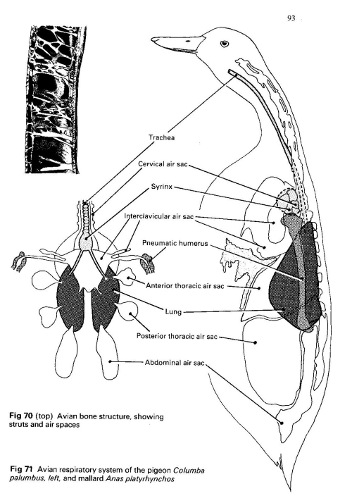
Drawing from How Birds Work by Ron Freethy
These 9 air sacs have very thin walls with few blood vessels, so they do not play a direct role in gas exchange to the body tissues. The gas exchange between the ventilation system and the blood which transports the oxygen to the body cells takes place in the air capillaries which rise up from the Para bronchi. The air sacs act as a bellows to ventilate the lungs. (Powell 2000) The avian respiratory system uses "flow-through ventilation” by relying on the flexible air sacs to move the inhaled air through the rigid lungs. Air passes thru the lungs on both the inhalation and exhalation phases. The 9 air-sacs greatly enhance the efficiency of the system and allow for the high metabolic rate found in birds.
It actually takes 2 inhalations to totally exchange the air within the avian respiratory system. During the first inhalation, air enters through either the nares or the mouth, passes through the pharynx and down the trachea directly into the posterior air sacs, while simultaneously into the lungs and all the way to the Para bronchi and into the anterior air sacs. During the exhalation, air moves out of the posterior air sacs, into and through the Para bronchi and, simultaneously, out of the anterior air sacs and out of the body via the trachea. The 2nd inhalation will bring in a new supply of fresh air. This unidirectional flow means that air moving through the lungs of a bird is largely ‘fresh’ air and has a higher oxygen content than air coming into the lungs of mammals which is mixed with air that has been in the lungs for a while.
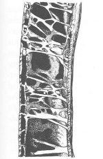 Oxygen Storage Pockets
A portion of the air sac system actually penetrates the skeleton at the humerus, vertebrae and femur, forming air pockets in otherwise dense bone. The exact reason that this function evolved is not known but it is theorized that it happened to lighten the bone structure, allowing dinosaurs to walk upright and the ancestor of birds to fly. These oxygen storage cavities make it possible for birds to fly long distances (during migration) at high altitudes where oxygen is in short supply.
Help to maintain body warmth
Now that
you can see just a small part of how important the
ventilation/respiratory system is in your Gouldian Finches, it might be
easier to understand that when they are infested with air sac mites it  will
affect everything that they do. Since the ventilation system helps to
maintain their body temperature system, when it is not functioning
properly your birds will become fluffed and frumpy in an attempt to
control their body temperature by external means. They are attempting to
create an insulation blanket around themselves like we would when we
cover up with heavier clothing during the colder months of the year.
will
affect everything that they do. Since the ventilation system helps to
maintain their body temperature system, when it is not functioning
properly your birds will become fluffed and frumpy in an attempt to
control their body temperature by external means. They are attempting to
create an insulation blanket around themselves like we would when we
cover up with heavier clothing during the colder months of the year.
The best course of action when you see this behavior in your birds, whether the cause is air sac mites or some other illness causing the bird distress, would be to supply them with an external heat source to help maintain a normal body temperature so that the bird does not have to use what energy it is able to produce to keep itself warm, but can use that energy to help itself heal. The external heating should not come from a source that also provides light, such as an electric light bulb. You should use something that provides only heat, such as a ceramic reptile heater, an infrared heat bulb, or even a heated perch system. A warmed hospital cage can be fashioned quite inexpensively by using a heating pad under a small cage that has one perch hanging very near the bottom of the cage to facilitate the bird absorbing as much warmth as possible radiating up through the floor of the cage. Place white paper toweling on the floor and be sure to locate food and water near the perch. A partial covering over the cage with a cloth will keep the warmth contained where it is needed. One important note: only older versions of a heating pad can be used in this manner, because our government has taken it upon itself to "protect” us from ourselves and now require that all newly manufactured heating pads automatically shut off after 2 hours.
Air Sac Mite studies
In 1992, Sonia C. Tidemann et al. ran extensive studies on the relationship of the rhinonyssid parasite Sternostoma tracheacolum (air sac mite) and the Gouldian Finch. Her goals were to:
- Determine the prevalence and intensity of infection in the wild Gouldian Finches compared to other finch species living in the same northern territory of Australia
- Determine the earliest date of occurrence in wild birds in Australia
- Determine the pathological effect of S. tracheacolum on individual birds.
Prevalence and Intensity of Infection
These studies found air sac mites in Gouldian Finches and 6 other species living in close proximity to them. The other 6 species were Long-tailed Finches, Masked Grassfinches, Pictorella Manikins, Zebra Finches, Double-barred (Owl) Finches, and Budgerigars. But the prevalence and intensity of infection in Gouldian Finches was higher than in the other species except the Pictorella Manikins.
Timetable for Infection
The studies were unable to determine the exact date that the air sac mites arrived in Australia because they were unable to obtain any wild bird specimens (whole birds) from museums anywhere in the world housing collections of Australian avifauna collected on early, historical expeditions. It was supposed that the introduction of the air sac mite into Australia may have been recent. The source possibly being migratory birds or aviary birds that came into Australia before quarantine procedures were mandatory. Fain and Hyland (1962) suggest that the normal hosts of S. tracheacolum in the northern hemisphere are wild birds because infected wild birds survive better than infected captive Canaries.
Pathological Effect
(This is the beginning of the technical stuff, so don't stop reading now)S. tracheacolum is a virulent species of nasal mite known to enter the trachea, lungs and even the body cavity of perching birds throughout the world (Baker et al. 1956; Fain and Hyland 1962). This mite causes inflammation, lesions, parasitic nodules, and hemorrhaging of the respiratory system leading to pneumonia and ultimately death of Canaries and the Gouldian Finch (Baker et al. 1956; Cumming 1959).
In the Tidemann study, the lungs of infected birds contained disintegration of the tissue lining the site of mite attachment as well as the infiltration of macrophages and lymphocytes to fight off the invading mites. The air sacs were thickened by excess fluid accumulation and infiltration of fibroblasts and lymphocytes. The trachea also had moderate epithelial lesions and lymphocyte and macrophage infiltration, again to fight off the invading parasites.
The mites were 0.2 – 0.3mm wide, 0.4 - 0.6mm long and dorso-ventrally flattened. Scanning electron micrographs indicated that individuals attached themselves by embedding their legs into connective tissue. A mucus coat, secreted by the bird’s immune system covered most of the individual mites that they found attached to the lining of the respiratory system. When mites were found in the trachea, they (plus their mucus coats) reduced the internal volume of the trachea. So you can see why it is hard for infected Gouldians to breathe once the width of the trachea is reduced and they are unable to get enough air to pass on down into their ventilation system. As the infestation of mites grows, the trachea narrows even further, and the birds will begin breathing with an open beak in an attempt to bring more air into their respiratory system. This same mucus coating will also thicken the wall of the air sac at the site of the mite attachment, thus preventing the air sac from acting as an efficient bellows to move air in and out of the lungs. The harder their breaths become the more bodily movement you will notice. This explains why "tail bobbing” is often stated as a symptom of an air sac mite infection. A healthy finch without an air sac mite infection will not noticeably move when they are breathing.
Immune System Checks
In the Tidemann study, there was a 62% infection rate in the Gouldians studied, so it does seem that it is possible to have Gouldian Finches that have never been infected with air sac mites. Through the centuries, in the face of continuous threats from parasites, birds have developed an elaborate system of preventive and controlling measures to keep themselves free of invading organisms – this elaborate system is a quality, high functioning immune system. In our captive Gouldians, air sac mite infections are a rare event when adequate nutrition is present to allow the birds to develop a strong immune system from time of hatch. So even if a few mites invade the respiratory system, the immune system will keep their numbers in check, preventing a life threatening infestation.
But stress always lowers the immune response in all living organisms. Most of our captive finches are under a constant amount of stress, if not from their molting cycle, breeding cycle or crowded living conditions then just from the fact that they are captive and living behind bars. I know what you are going to say now…”but they have always lived in a cage, they have never known anything else”. But have you ever watched your flock as you approach them on a daily basis. They see you every day of their lives. You feed them and clean up after them. Yet, if they are all down eating at their food dish, and you approach, they will fly up in unison to higher perches and a position of safety. Whenever this fight or flight mechanism is triggered, there is resulting stress, if only for a very short time period.
Transmission Biology Studies
Studies
conducted in 1995 by P. J. Bell explored the parasite-host relationship
and the transmission biology of the air sac mites to the Gouldian Finch.
These studies found that S. tracheacolum was
ovoviviparous (producing eggs that develop within the maternal body and
hatch within or immediately after release from the parent body). The
young are laid in the lung of the host (your birds) and molt 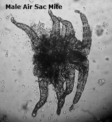 without feeding. Following a blood meal the female protonymphs move to the posterior air sacs (where
they are protected from the host’s immune response because there is
little blood flow to the air sacs preventing any insecticide in the
bloodstream during this time from reaching them) The male
protonymphs tend to stay within the lungs, where there is significant
blood flow, to complete development and therefore can be killed when an
insecticide is present in the blood stream. Pregnant
females tend to occupy the air sacs, the syrinx and trachea of the host
while adult non-pregnant females are more commonly found in the upper
respiratory system, particularly the mouth and nasal cavities. In vitro rearing experiments and other observations indicate that the life cycle (from egg to adult) is completed in less than 6 days.
without feeding. Following a blood meal the female protonymphs move to the posterior air sacs (where
they are protected from the host’s immune response because there is
little blood flow to the air sacs preventing any insecticide in the
bloodstream during this time from reaching them) The male
protonymphs tend to stay within the lungs, where there is significant
blood flow, to complete development and therefore can be killed when an
insecticide is present in the blood stream. Pregnant
females tend to occupy the air sacs, the syrinx and trachea of the host
while adult non-pregnant females are more commonly found in the upper
respiratory system, particularly the mouth and nasal cavities. In vitro rearing experiments and other observations indicate that the life cycle (from egg to adult) is completed in less than 6 days.
Adult male mites tend to be common in small populations but uncommon and often absent in large populations, supporting an argument for parthenogenesis (biological reproduction that involves development of a female sex cell without fertilization) and an arrhenotokous (producing only males) system of sex determination.
Transmission from one bird to another is accomplished by the adult non-pregnant females as this stage was observed on the head plumage, beak and nares of infected birds. Artificial infection experiments indicated that non-pregnant females from the nares of Gouldian Finches are capable of surviving long enough to start a new infection in a naive host. Bell’s observations indicated that adult non-pregnant females possessed suitability for prolonged exposure to ambient conditions, an attribute not found in other life stages of the air sac mite. Further observations indicated the potential for these non-pregnant females to remain on the bill and plumage for many hours before retreating to the humid safety of the nasal cavity. These females were regularly observed on the external nares, mandibles and head plumage of infected Gouldian Finches. These female mites were observed to carry the first pair of legs in the air and wave them vigorously. These adult non-pregnant females were observed in all months of the year, between 08.00 and 18.00 hours and on both male and female birds in both breeding and non-breeding condition.
Bell’s study stated that S. tracheacolum is not a passive participant in the process of transmission but has the potential to actively seek a new host. He felt that it was unlikely that transmission occurred as regurgitant between parent birds or between parent and chick as the mechanical action of this activity alone may be sufficient to destroy the delicate mites living inside of the birds. He stated that any mite surviving transferral in this manner to the crop of a nestling bird must then survive further mechanical and biochemical dangers during migration to the trachea.
The Bell studies clearly state that they felt that the most probable modes of transfer were (1) direct from the nasal cavity of an infected bird to the nasal cavity of a recipient bird via the external nares of each bird and (2) indirect from the nasal cavity of an infected bird to the nasal cavity of a recipient bird via the external nares, but including a period of time on water, perches or nest material. This is why it is very important that you thoroughly clean and disinfect the cages and nestboxes if any are present during treatment for air sac mite infestations.
Posterior Air Sac protection
Once a bird is infected, it is next to impossible to ever completely eradicate them from a birds respiratory system because the female protonymphs migrate to the posterior air sac where they are protected from the bird’s immune system and from the insecticides that we have available to us that will kill the adult form of the air sac mite. Remember, there is little blood flow in the air sacs themselves, so it is impossible to get the insecticide that is flowing in the bird's blood stream to the protonymphs living in the air sacs. A bird with a strong, healthy immune system can keep these nymphs in dormancy for many months, but when the bird's immune system is suppressed during times of stress, like breeding and molting, the nymphs will eventually mature and begin the whole life cycle again.
When a bird has a strong, healthy immune system it is able to keep the population of nymph air sac mites from maturing. During very stressful times of the year (breeding and molting seasons), when the immune system becomes suppressed it is up to us to help them keep the adult numbers in check as the protected nymphs mature. We can do this by using either Ivermectin (S76) or Moxidectin (Scatt). If we fail to do this, before the population of mites as grown out of control, the simple act of treating the bird to kill the mites could likely also suffocate the bird itself.
Treatments to kill air sac mites
Since Scatt remains in a bird's blood stream for 3 weeks, I do not like using it more than 4 times per year…immediately prior to and immediately after both the breeding season and molting cycle. S76 will only remain in the blood stream while it is available to the bird in its drinking water, so the recommendation is that during the molting and breeding cycles the treatments should be given for 2 consecutive days every 3 weeks. The rest of the year it should be administered for 2 consecutive days every month.
ПРОДОЛЖЕНИЕ: http://amadinagoulda.ru/index/why_all_the_fuss_about_air_sac_mites_ii/0-283